CT Scan (Computed Tomography Scan), also known as CAT (Computed Axial Tomography) scan, is a common medical imaging technique that uses X-rays and computer processing to create detailed cross-sectional (slice-like) images of the body.
What is a CT Scan?
- CT scan takes hundreds or thousands of X-ray measurements from many different angles around the body.
- A computer then reconstructs these into thin “slices” that can be viewed individually or form 3D-like images.
- This allows doctors to see internal structures with much greater detail and clarity than standard X-rays
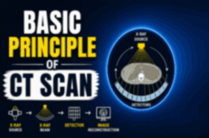
How Does a CT Scan Work? (Basic Principle)
- X-ray Source & Rotation: The patient lies on a movable table that moves through a doughnut-shaped machine (called a gantry). Inside the gantry, an X-ray tube rotates rapidly around the patient (often in a spiral/helical path in modern scanners), sending narrow beams of X-rays through the body.
- Detectors: On the opposite side, detectors measure how much the X-rays are attenuated (weakened or absorbed) as they pass through different tissues.
Computer Processing:
- Dense tissues (like bone) absorb more X-rays → appear white.
- Less dense tissues (like air in lungs) absorb less → appear darker.
- Soft tissues show varying shades of gray.
- The computer uses mathematical algorithms (tomographic reconstruction) to create the final images.
- Hounsfield Units (HU): Each pixel in the CT image is assigned a number based on tissue density (e.g., bone is high, water is 0, air is negative).
Common Uses
- Head: Detect tumors, bleeding, strokes, injuries.
- Chest: Lung cancer, pneumonia, blood clots (pulmonary embolism).
- Abdomen/Pelvis: Kidney stones, appendicitis, liver issues, cancers.
- Bones & Spine: Fractures, spinal problems.
- Blood vessels (with contrast): Angiography.
- Guiding biopsies, planning surgery or radiation therapy.
- Contrast dye (iodine-based, injected or swallowed) is often used to highlight blood vessels or organs better.
Procedure –
- Usually takes 10–30 minutes.
- You lie still on the table; it slides through the scanner.
- You may be asked to hold your breath briefly.
- The machine is open (not fully enclosed like some MRIs),
- No pain, but contrast can cause a warm sensation or metallic taste.
Advantages
- Fast (much quicker than MRI).
- Excellent for bones, lungs, and detecting acute issues like bleeding or trauma.
- Widely available and relatively affordable.
- Can produce 3D reconstructions.
Disadvantages & Risks
- Radiation: Involves ionizing radiation (higher dose than a regular X-ray, typically 1–10 mSv depending on the area). Doctors follow “ALARA” (As Low As Reasonably Achievable) principles.
- Risk of allergic reaction to contrast (rare).
- Not as good as MRI for soft tissue detail in some cases (e.g., brain or joints).
CT Scan
- Best for Bones, lungs, emergencies,Soft tissues, brain, joints
- Time,Fast (5-10minutes)Longer (20–60+ min)
Cost/Availability
- Generally cheaper & faster
- More expensive
- CT scans are a vital tool in modern medicine, especially in emergencies. Always discuss benefits vs. risks with your doctor, especially if pregnant or concerned about radiation.
I hope guys like this post Basic Principle of CT Scan